2026-09-22
Dual Canthoplasty (Lateral & Lower Canthoplasty): Surgical Techniques and Precautions
Dual canthoplasty combines lateral and lower canthoplasty to horizontally lengthen and vertically soften the outer eye contour. Learn the techniques, precautions, and recovery process.
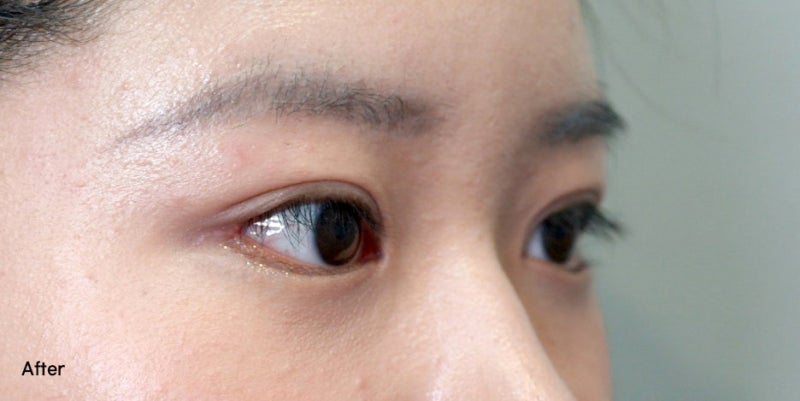
Dual Canthoplasty (Lateral & Lower Canthoplasty): Surgical Techniques and Precautions
The eyes play a critical role in shaping a person's first impression. Because of this, many people who are dissatisfied with the size, shape, or slant of their eyes consider aesthetic changes.
Depending on factors such as skin laxity, eyelid thickness, and unique eye characteristics, different procedures may be indicated, including double eyelid surgery, ptosis correction, or canthoplasty.
In particular, individuals whose eyes appear closed-off due to a short horizontal length, or sharp and fierce due to an upward-slanting outer corner, may consider surgical approaches addressing the lateral canthus.
Today, we will examine dual canthoplasty—which addresses both the horizontal and vertical dimensions of the outer eye by combining lateral and lower canthoplasty—including its anatomical principles and critical precautions.
Concept and Characteristics of Dual Canthoplasty
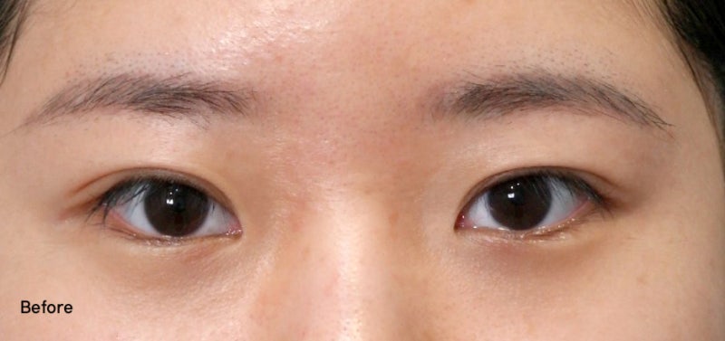
Every individual has unique eye anatomy, orbital bone depth, and scleral exposure (the visible white of the eye). Even with a well-defined double eyelid crease, an elevated lateral canthus or a narrow outer margin can create a harsh, sharp appearance.
Canthoplasty targeting the lateral canthus is one effective way to modify this structural contour.
Commonly referred to as dual canthoplasty, this technique synergistically combines lateral canthoplasty—which extends the outer horizontal length—with lower canthoplasty—which lowers the outer contour of the lower eyelid.
Synergistic Mechanism of Lateral and Lower Canthoplasty
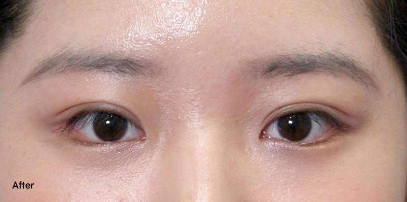
When widening the eye, pulling tissue in only one direction can result in an unnatural curvature or cause the opening to narrow again due to tissue adhesions. Therefore, both directional vectors must be harmoniously integrated.
Lateral Canthoplasty: Involves delicate dissection of the outer edge of the eye (lateral canthus) and secure fixation to the periosteum. The primary objective is to lengthen the horizontal width and achieve balanced facial proportions. Internal tissues are precisely dissected to prevent excessive conjunctival exposure (eversion), and specialized anchoring methods are applied to minimize the risk of the corners re-adhering.
Lower Canthoplasty: When the lower eyelid covers too much of the outer iris, resulting in a confined or sharp look, this approach accesses tissue transconjunctivally (through the inner eyelid) to lower the lower lid along a gentle, diagonal curve. By eliminating external skin incisions, it leaves no visible scars on the surface while creating a softer, more natural contour.
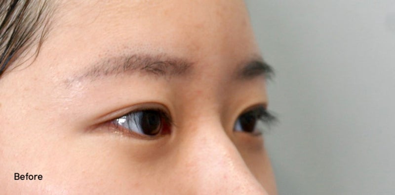
To put it simply, imagine a narrow window: widening the side frame outward while lowering the bottom sill allows significantly more light and visibility to enter in a balanced manner.
This method safely increases the exposure of the outer sclera, creating a softer, gently curved outer eye angle.
Avoiding Overcorrection and Understanding Anatomical Limits
In dual canthoplasty, aggressively widening the eyes does not guarantee optimal outcomes.
Each patient has defined anatomical boundaries for safe tissue relocation, determined by orbital bone structure, conjunctival laxity, and the degree of ocular prominence.
If excessive resection or over-traction is performed without accounting for individual anatomy, the following complications may occur:
◾ Deformity of the lateral canthal angle into an unnatural rounded or squared 'C' or 'U' shape
◾ Excessive outward exposure of the conjunctiva or red mucosal tissue (ectropion)
◾ Functional issues such as dry eye syndrome, persistent tearing (epiphora), or ocular sensitivity
Consequently, the surgeon must accurately identify the lateral canthal structures and periosteal anchoring points, employing meticulous surgical design tailored to individual anatomy to minimize tissue trauma.
Recovery Process and Potential Side Effects
Following canthoplasty, temporary swelling, bruising, chemosis (conjunctival swelling), and subconjunctival hemorrhage may occur. These symptoms typically subside gradually as tissues stabilize, though recovery rates vary by individual.
· Avoid rubbing, pressing, or applying mechanical force to the eyes during early recovery.
· Postpone wearing contact lenses and applying eye makeup until the conjunctiva and incision sites have healed completely.
· Depending on individual factors, risks such as bleeding, infection, asymmetry, scarring, and ectropion may arise; close follow-up and proper post-operative care are required.
Importance of Specialist Diagnosis and Consultation
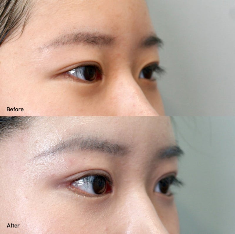
Rather than applying a standardized, one-size-fits-all surgical technique, successful eyelid surgery relies on a thorough analysis of overall facial proportions and orbital bone anatomy.
Because skin elasticity, scar response, recovery timelines, and anticipated results differ from person to person, it is strongly advised to undergo a comprehensive consultation with a board-certified plastic surgeon to formulate a safe, tailored surgical plan.










Frequently Asked Questions
What is dual canthoplasty (lateral and lower canthoplasty)?
It is a combined surgical procedure that simultaneously performs lateral canthoplasty (lengthening the outer horizontal width) and lower canthoplasty (lowering the outer lower eyelid margin). By expanding both horizontal and vertical dimensions, it exposes more of the outer sclera and softens the angle of the eye.
Does lower canthoplasty leave visible external scars?
No, external scarring is minimal. Lower canthoplasty is typically performed transconjunctivally from the inner side of the eyelid rather than through external skin incisions, significantly reducing the risk of visible scars while lowering the lower eyelid in a gentle diagonal slope.
What complications can arise from overcorrecting canthoplasty?
Complications can include canthal angle deformities and excessive mucosal exposure. Ignoring the anatomical boundaries of the orbit and conjunctiva may cause the outer corner to become rounded or webbed into a 'C' or 'U' shape, expose the red conjunctiva, or trigger functional issues like severe dry eye, tearing, and stinging sensations.
What post-operative care is essential after dual canthoplasty?
Patients should avoid rubbing, pressing, or straining the eye area during initial healing. Contact lenses and eye makeup must be avoided until the incisions and conjunctiva have completely healed, and ample rest is recommended while temporary swelling, bruising, and chemosis gradually resolve.
Why does the outer corner sometimes re-adhere or narrow after surgery?
Re-narrowing can occur if tissues are pulled along a single vector or if post-surgical adhesions develop. To prevent this, surgeons precisely dissect the lateral canthal tissues, securely anchor them to the periosteum, and harmoniously coordinate the lower eyelid vector to minimize tissue re-adhesion.